Microwave Tomography Explained Current State of the Art and Why It Is Safe

The word “microwave” can make a medical imaging technology sound more alarming than it is. Most people hear it and picture a kitchen appliance heating leftovers. Microwave tomography uses the same broad part of the electromagnetic spectrum, but in a very different way: low-power signals, carefully measured, used to build a map of what is inside an object or body.
That distinction matters. Microwave tomography is being studied for breast imaging, stroke assessment, lung monitoring, industrial inspection, and other uses where safe, repeatable imaging could be valuable. It does not replace CT, MRI, ultrasound, or X-ray in everyday care today. It is still a developing field. But the science behind it is solid enough that researchers, engineers, and clinicians continue to push it forward.
This article explains how microwave tomography works, where the technology stands now, and why the safety profile is far less scary than the name suggests.

What microwave tomography actually measures
Microwave tomography is an imaging method that sends very low-power microwave signals into an object and measures how those signals scatter, slow down, weaken, or change direction.
Different materials interact with microwaves in different ways. In the body, tissues vary in two key electrical properties:
Permittivity
How strongly a material stores electrical energy from an electromagnetic field.
Conductivity
How easily electrical current can move through the material.
Water-rich tissue, fatty tissue, blood, air-filled regions, and abnormal tissue can all produce different microwave responses. A microwave tomography system measures those responses from many angles, then uses mathematical reconstruction to estimate the internal structure.
The result is not a photograph. It is more like a property map. Instead of showing anatomy in the same way as an MRI scan, microwave tomography estimates the electrical characteristics inside the scanned region.
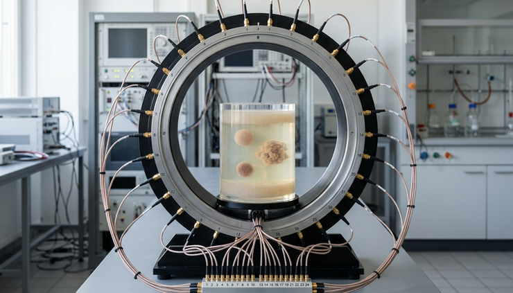
A simple system usually includes:
A set of small antennas placed around the area being scanned
A signal source that emits controlled microwave energy
Sensors that record the signals after they pass through or scatter
Software that reconstructs an image from the measurements
This process is challenging because microwaves do not travel through tissue in straight lines. They scatter, reflect, and interact with many boundaries. That makes the image reconstruction problem much harder than basic X-ray imaging. It is one reason the field has taken years to mature.
Why this is not the same as putting tissue in a microwave oven
A kitchen microwave oven is designed to heat food. It uses relatively high power and traps energy inside a metal cavity so water molecules in food absorb that energy and warm up.
Microwave tomography does something very different.
It uses low-power diagnostic signals, not cooking-level energy. The goal is to measure how the signal changes, not to deposit enough energy to heat tissue. In many research and prototype systems, the emitted power is comparable to or below levels used by common wireless technologies, depending on the design and frequency.
The key difference is intent and exposure:
Kitchen microwave oven | Microwave tomography |
Designed to heat food | Designed to measure signal changes |
Uses high power inside a closed chamber | Uses low-power signals under controlled conditions |
Energy absorption is the point | Heating is avoided and monitored |
Not an imaging tool | Built specifically for sensing and reconstruction |
Microwaves are a form of non-ionizing radiation. That means they do not carry enough photon energy to break chemical bonds or directly damage DNA in the way ionizing radiation can. X-rays and CT scans use ionizing radiation. Microwave tomography does not.
That does not mean any microwave exposure is harmless at any level. High enough power can heat tissue. Safety depends on frequency, power, duration, antenna design, and how much energy the body absorbs. But diagnostic microwave tomography is designed around low exposure and safety limits.
The safety science behind microwave tomography
The main known biological effect of microwave energy at sufficient intensity is heating. Safety engineering focuses on keeping absorbed energy low enough that meaningful tissue heating does not occur.
Researchers and device developers look at several factors.
Specific absorption rate is central
Specific absorption rate, often called SAR, describes how much electromagnetic energy tissue absorbs per unit mass. It is also used in safety discussions around mobile phones and MRI radiofrequency fields.
A microwave tomography device must be designed so exposure stays within accepted safety limits for the intended use. That involves:
Controlling output power
Limiting scan duration
Using antenna designs that distribute energy safely
Modeling energy absorption before human testing
Measuring device performance under realistic conditions
Regulated medical devices must also go through safety testing before clinical use. The exact requirements depend on the country, device type, and intended application.
Low power reduces heating risk
Because tomography depends on measuring scattered signals rather than forcing energy into tissue, the system can operate at low power. More power is not automatically better. Too much energy can distort measurements, create safety issues, and complicate the reconstruction.
Engineers usually want the weakest signal that still gives useful data.
Non-ionizing does not mean unregulated
The term “non-ionizing” sometimes gets misunderstood. It does not give any device a free pass. It only describes the type of radiation and its interaction with matter.
A safe microwave tomography system still needs:
Careful exposure design
Hardware safeguards
Thermal and electromagnetic modeling
Quality control
Clinical validation for medical claims
So the right safety message is balanced: microwave tomography avoids the DNA-damaging mechanism associated with ionizing radiation, and its power levels are designed to avoid heating. It still must be tested and regulated like other medical technologies.

Where the technology is most promising
Microwave tomography is attractive because it can be safe, relatively low cost, portable, and repeatable. Those strengths matter most in situations where repeated imaging is useful or where large scanners are hard to access.
Breast imaging
Breast imaging has long been one of the most studied medical uses. Breast tissue often has strong contrasts in electrical properties, especially between fatty and water-rich tissue. That makes it a natural target for microwave methods.
Researchers have explored microwave tomography as a possible adjunct to mammography, ultrasound, or MRI. It is not a routine replacement for mammograms. Mammography remains a proven screening method with established clinical workflows.
The appeal of microwave breast imaging is that it may offer a non-ionizing, comfortable, repeatable way to gather additional tissue information. Some systems use a cup or array design around the breast, with antennas arranged to collect data from many directions.
The challenge is accuracy. Breast anatomy varies widely. Tissue composition differs from person to person. Small lesions can be hard to reconstruct clearly. This is why clinical validation remains essential.
Stroke and brain monitoring
Microwave systems are also being studied for detecting or monitoring stroke, especially because blood and brain tissue can have different electrical responses.
A portable system that helps distinguish bleeding from blockage would be valuable in emergency care, but this is a hard problem. The skull affects microwave propagation. Brain structures are complex. Motion, anatomy, and patient-to-patient differences all complicate the signal.
This area remains active in research. The promise is real, but routine clinical use requires evidence that the method works reliably across many patients and emergency conditions.
Lung and chest monitoring
The lungs contain air, fluid, blood, and tissue, all of which affect microwave signals differently. That makes chest monitoring another area of interest.
Potential uses include tracking fluid changes or monitoring respiratory conditions, but the chest is a complex imaging target. The ribs, heart, lungs, and movement from breathing all influence measurements.
Microwave tomography may be most useful here as a monitoring tool rather than a one-time diagnostic image. Repeated safe measurements could show trends over time.
Industrial and security inspection
Not every use is medical. Microwave tomography can also inspect materials, detect moisture, find voids, or evaluate structures that respond differently to microwave signals.
For example, it may help identify water content in materials, inspect composites, or detect hidden defects. These uses are often easier than medical imaging because the object is more controlled and does not move, breathe, or vary biologically.
The current state of the art
Microwave tomography has moved far beyond simple lab demonstrations, but it is not yet a mainstream medical imaging tool. The current state is best described as technically advanced, clinically selective, and still maturing.
Several trends define the field today.
Better antennas and measurement hardware
Modern systems use improved antenna arrays, more stable electronics, and better calibration methods. This matters because microwave tomography is sensitive to small errors. A slight mismatch in antenna response or a small positioning error can affect the reconstructed image.
New hardware aims to collect cleaner data from more angles in less time. That can improve resolution and reduce artifacts.
Stronger reconstruction algorithms
The hardest part of microwave tomography is turning scattered electromagnetic measurements into a useful image. This is an inverse problem, which means the system works backward from measured signals to estimate what internal structure caused them.
That problem is mathematically difficult. Many possible tissue maps could explain similar measurements. Good algorithms use physics, constraints, prior information, and repeated calculations to find the most likely image.
Recent work often combines electromagnetic modeling with machine learning. Machine learning can help speed reconstruction, reduce noise, or improve pattern recognition. Still, it must be trained and tested carefully. A model that works well on simulated data may struggle with real human variation.
Hybrid imaging approaches
Some research systems combine microwave data with other imaging methods. For example, ultrasound or MRI geometry can guide microwave reconstruction. This can reduce uncertainty because the algorithm starts with better information about the body’s shape and tissue boundaries.
Hybrid approaches may become important because microwave tomography alone can face limits in spatial resolution. Pairing it with another modality can help each method do what it does best.
More realistic testing
Early imaging studies often used simple phantoms, such as containers filled with tissue-like materials. Those are still useful, but the field has increasingly moved toward more realistic phantoms, human studies, and application-specific testing.
That shift is important. A system can perform well in a controlled lab setup and still struggle in clinical use. Real patients move. Bodies vary. Skin contact changes. Temperature changes. Cables shift. Clinical workflows are messy.
The state of the art now focuses less on proving the basic concept and more on proving reliability.
What still limits microwave tomography
The main limits are not safety. They are image quality, validation, and clinical fit.
Resolution is different from CT or MRI
Microwave wavelengths are larger than X-ray wavelengths, which affects spatial resolution. Microwave tomography often cannot show tiny anatomical details the way MRI or CT can.
That does not make it useless. Many tools measure things that other tools do not. A thermometer does not show anatomy, but it can still be medically important. Microwave tomography may succeed where electrical property contrast is more useful than fine structure.
Tissue complexity makes reconstruction hard
Human tissue is layered, irregular, and variable. Fat, muscle, bone, blood, fluid, and skin all affect signals. The more complex the region, the harder the image reconstruction.
Good calibration helps. Better models help. Large clinical datasets help. But this remains one of the biggest barriers.
Clinical adoption needs proof
Hospitals and clinics do not adopt imaging tools just because they are safe. A device also needs to answer a real clinical question better, faster, cheaper, or more comfortably than existing options.
That proof takes time. Researchers must show that microwave tomography can produce reliable results, fit into workflows, and improve decision-making. For some applications, it may become an add-on rather than a replacement.
Why the word microwave should not scare people away
The fear around the term comes from a reasonable place. People know microwaves can heat things. But safety depends on dose, power, duration, and device design.
Visible light can be harmless from a lamp and dangerous from a high-power laser. Sound can be pleasant through headphones and harmful at extreme volume. The same broad category of energy can have very different effects depending on how it is used.
Microwave tomography uses controlled electromagnetic signals to gather information. It is closer in spirit to radar or wireless sensing than to cooking food.
The main safety points are clear:
It uses non-ionizing radiation
It is designed to avoid harmful heating
It can operate at low power
Exposure can be modeled, measured, and limited
Medical use requires safety testing and regulatory review
For patients, the practical question is not “Are microwaves always safe?” The better question is “Has this specific device been tested for this specific use?” That is the same standard that should apply to any medical technology.
How it compares with other imaging methods
Each imaging method has strengths and tradeoffs.
X-ray and CT provide high-resolution structural images, but they use ionizing radiation. MRI provides detailed soft-tissue images without ionizing radiation, but scanners are large, expensive, and not suitable for every patient. Ultrasound is portable and safe, but image quality depends heavily on anatomy and operator skill.
Microwave tomography may offer:
Lower-cost hardware compared with large scanners
Portable or bedside designs
Repeatable measurements without ionizing radiation
Sensitivity to tissue electrical properties
Potential use in monitoring over time
It may struggle with:
Fine spatial detail
Complex anatomy
Standardization across patients
Long validation pathways
Competing with established tools that already work well
The likely future is not a world where microwave tomography replaces every other scanner. A more realistic future is one where it fills specific gaps.
What the next few years may bring
Progress will likely come from focused applications rather than broad promises. The most successful systems will probably target a specific question, such as detecting a meaningful tissue change, monitoring a known condition, or helping triage a narrow clinical problem.
Expect work in several areas:
Faster scans with better antenna arrays
Improved calibration for real-world use
Physics-guided machine learning
Larger clinical studies
More portable systems
Better safety and quality standards for specific applications
The technology will also benefit from advances in computing. Microwave reconstruction can require heavy calculations. Faster processors and better algorithms make real-time or near-real-time imaging more practical.
But the field still needs patience. Medical imaging technologies earn trust through repeated evidence, not excitement alone.

The takeaway
Microwave tomography is a real imaging and sensing technology built on low-power electromagnetic measurements. It maps electrical properties inside tissue or materials, which can reveal information that other imaging methods may miss.
Its safety profile is one of its strengths. The signals are non-ionizing, the power levels are controlled, and the goal is measurement rather than heating. The word “microwave” may sound unsettling, but a diagnostic microwave tomography system is not a microwave oven pointed at the body.
The honest view is both optimistic and careful. The technology is promising, especially for portable and repeatable imaging, but it still faces hard challenges in resolution, reconstruction, and clinical proof. If continued research shows that it can answer specific medical questions reliably, microwave tomography could become a useful companion to today’s imaging tools.
This article is informational only and is not medical advice. For any imaging decision, the right source is a qualified clinician using an approved device for a specific medical need.


Comments